Friday 12 June 2015

Gross and microscopic structure of skeletal muscle. The ultrastructure of a myofibril. The roles of actin, myosin, calcium ions and ATP in myofibril contraction. The roles of calcium ions and tropomyosin in the cycle of actinomyosin bridge formation.

The roles of actin, myosin, calcium ions and ATP in myofibril contraction. The roles of calcium ions and tropomyosin in the cycle of actinomyosin bridge formation.

In a muscle cells join up to make muscle fibres, resulting in long strands of cells sharing nuclei and cytoplasm (aka sarcoplasm). Also inside these fibres are myofibrils: these are the structures that allow contraction. Myofibrils are made up of proteins.

Fibers are grouped together into bundles which are grouped further to create muscles.


Myofibrils are made from myofilaments. These are proteins which mean contraction can happen. They are arranged in such a ways as to create the appearance of different coloured bands: A-band and I-band (easily remembered by thing dArk band and lIght band). The distance between two z-lines is called a sarcomere.

(turned 90 degrees from previous diagram)
The two filaments are called actin and myosin.
Actin

Myosin
During contraction, the heads of myosin go into the dips in actin and then push it so that the two are propelled in opposite directions. This makes them move like they are crawling up one another, bringing the z-lines closer together and decreasing the size of myofibrils and therefore muscles overall.

For this to happen, Ca2+ binds to troponin, which changes its positioning moving tropomyosin out of the binding site of actin.
This allows myosin heads to bind there, as they do they change angle and release a ADP molecule. ATP then attatches to the head causing it to detach from the actin. The ATP turns to ADP providing energy for the head to return to where it was before so that it can repeat the process.

An organism’s genome contains many repetitive, non-coding base sequences. The probability of two individuals having the same repetitive sequences is very low. The technique of genetic fingerprinting in analysing DNA fragments, that have been cloned by PCR, and its use in determining genetic relationships and in determining the genetic variability within a population. Candidates should be able to - explain the biological principles that underpin genetic fingerprinting techniques - interpret data showing the results of gel electrophoresis to seperate DNA fragments - explain why scientists might use genetic fingerprints in the field of forensic science, medical diagnosis, animal and plant breeding.

Introns (parts of DNA that don't code for anything) are made up of repeating units called core sequences. Because every individual (apart from identical twins) had unique DNA, it is most likely that no two people will have the same core sequences.
 
This is the process of genetic fingerprinting:
  • Extraction: and then replication using PCR
  • Digestion: by restriction endonucleases
  • Separation: by gel electrophoresis; then alkali used to separate into single strands
  • Southern blotting; the strands are transferred on to a nylon membrane by absorbent sheets drawing them up by capillary action; fixed with UV
  • Hybridisation: marked probes (radioactive or fluorescent) are then added at specific temperatures and PHs to bind to specific core sequences
  • Development: if the probes are radioactive the pattern is transferred to x-ray film
The film or gel will then show a unique pattern of lines corresponding to the DNA.
 
Uses:
  • Forensics: comparing the genetic fingerprint of a suspect and DNA from a crime scene to see if they are from the same person (probability of the results being the same but from different people is calculated)
  • Medical diagnosis: if done with genes patients DNA can be compared to that of someone with a disease to see how similar they are thus how likely they are to get it (e.g. Huntington's); DNA from pathogens from a patient can be compared with that of known ones to find out what it is
  • Breeding: to identify desirable or undesirable genes
  • Determining genetic variability: if DNA from different animals of the same species is very similar there is little genetic variability
  • Paternity testing: if you have the DNA of a mother next to the DNA of her child and the DNA of someone who may or may not be the father, everywhere the DNA of the child does not match with the mother, it will match with the father if he is the real one


Wednesday 10 June 2015

The use of labelled DNA probes and DNA hybridisation to locate specific genes. Once located, the base sequence of a gene can be determined by • restriction mapping • DNA sequencing. Many human diseases result from mutated genes or from genes that are useful in one context but not in another, e.g. sickle cell anaemia. DNA sequencing and the PCR are used to produce DNA probes that can be used to screen patients for clinically important genes. The use of this information in genetic counselling, e.g. for parents who are both carriers of defective genes and, in the case of oncogenes, in deciding the best course of treatment for cancers. Candidates should understand the principles of these methods. They should be aware that methods are continuously updated and automated.

DNA hybridisation
We need to find genes that are causing problems so that we can alter them. This is done by DNA hybridisation: DNA probe is made by making a sequence of DNA is made that is complimentary to the gene and is fluorescent or radioactive (32p), the patients DNA is separated into two strands, the probe will bind to the gene and fluoresce or show up on a photographic plate to show where it is.

DNA sequencing
This is how to find out the order of bases of DNA:

  • DNA is split into a single strand
  • DNA polymerase is added
  • Primers that are complimentary to the begining of the DNA are added (so that DNA polymerase has somewhere to start from)
  • Free nucleotides are added
  • One type terminator nucleotide is added (either C, T, A or G) that stops DNA polymerase when it is added to the chain; it will be radioactively marked (or more recently, fluoresce in a specific colour)
  • DNA polymerase will make many complimentary strands of DNA, they may be stopped at some point in their making by the terminator nucleotide
  • This results in many pieces of DNA of different lengths, each length corresponding to a place on the DNA where the base complimentary to the terminator nucleotide is
  • This is done in four tubes, each one using a different terminator molecule
  • So you will end up with a fragment that corresponds to each base on the sequence (however this cannot be seen yet)


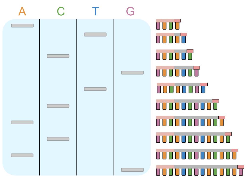
  • The mixture from each of the tubes is put on some agar gel, each in a separate column
  • A voltage is applied to the gel, which makes the far end positive, which attracts the negative DNA strands (gel electrophoresis)
  • Smaller molecule move more quickly and bigger molecules more slowly meaning that when the voltage is stopped the fragments are different distances through the agar base on size
  • Photographic film is put over the top and detects where each strand is because of the radioactive marking (or more recently the agar is scanned with lasers by computers)
  • This shows scientists the order of the complimentary bases so they can work out the sequence
Restriction mapping
DNA sequencing using the above method (sanger) can only be done using short fragments so DNA must be cut up using restriction endonucleases. Take for example this plasmid which has been cut up for sequencing:


Scientists cannot tell what order these fragments go in. In order to work it out they use the handy fact that restriction endonucleases only cut at specific sites.

To cut up this plasmid in the first place scientists would have used three restriction endonucleases, lets call them A, B and C. So, the whole plasmid is put in with just A and B and the lengths of the two resulting fragments recorded (using gel electrophoresis). This is then repeated but using just B and C, and then again using just A and C.

The whole plasmid is 10 kb (kilobases) long.
In the A and B trial there was one fragment of 2 and one of 8,
In the B and C trial one was 3 and the other was 7,
In the A and C trial there were two fragments of 5.

You can put all this information together to see that in between A and B must me 2 kb then in between B and C must be 3 kb and in between C and A must be 5 kb, because this adds up to 10. So now you can tell that the 2 kb fragment is first then the 3 kb fragment and then the 5kb fragment, and as the DNA for each of these fragments have been sequenced you can put it all together to know the sequence for the whole plasmid.


Sickle cell anaemia
This disease is caused by a substitution mutation on the gene for haemoglobin. It produces the amino acid valine instead of glutamic acid to make the protein haemoglobin s. Haemoglobin s has a sticky patch on it that makes it stick to other haemoglobin molecules making long, insoluble fibres (at low oxygen concentrations). This causes red blood cells to distort so they are sickle and inflexible meaning:

  • They block capillaries
  • They can't carry oxygen
  • Malaria cannot exist on them
The mutated and non mutated genes be haemoglobin are co-dominant. If an individual has two genes for haemoglobin s they will be very ill and possibly die from it. If one gene for haemoglobin s and one for normal haemoglobin (a) an individual will be mostly ok, unless their oxygen concentration gets particularly low.

Being heterozygous for this trait is useful in places with malaria, because it is difficult for the parasite to survive in your blood and you are not severely ill.

DNA screening
This is looking at peoples genes to see if they are carrying a harmful mutation. This is done by adding a marked fragment of DNA that is complimentary to the mutated gene (a DNA probe) to a strand of DNA from the patient, if the patient has the mutated gene the probe will bind to it and show up because it is radioactive.

Genetic counselling
This is advice given to people who have found out they have a harmful, or potentially harmful mutation in their genotype due to genetic screening. For example a couple will be told what the likely-hood is of their offspring having certain problems, what the implications of these problems are and how they will know in the future if their offspring have the problem or not.

There are many applications of this concerning cancer:
  • determining the best drugs to use to treat cancer (if the type of cancer is found out)
  • determining the risk of relapse (if the amount of mutations are looked at)
  • because two mutations are needed for a tumour to develop (oncogenes and inactive tumour suppressor cells) people may have one mutation, but not yet cancer, they can be told this to change their lifestyle in order to avoid the second mutation

Tuesday 9 June 2015

The use of gene therapy to supplement defective genes. Candidates should be able to evaluate the effectiveness of gene therapy.

Gene therapy is when a gene that is missing or not functioning is replaced by one that is.

There are two points at which gene therapy can happen:
  • Germ-line: where the genes are changed in a fertilised egg (could be a step towards eugenics)
  • Somatic-cell therapy: genes are changed in the affected cells of an adult (pretty ineffective as it must be repeated as cells die, hardly any of the DNA gets expressed and the DNA is not passed on)
There are two ways of changing genes:
  • The gene correct gene can be added to cells and a functioning protein will be produced because it is the dominant allele, this is gene supplementation.
  • The gene can be replaced altogether (the defective gene removed and the correct one put in).
There are two ways of delivering genes in somatic-cell therapy:
  • Using harmless transgenic viruses that inject their recombinant DNA into the affected cells (can create immune response and be rejected)
  • Putting the DNA in lipids which can cross the cell surface membrane (liposomes) (can create immune response and be rejected)
Gene therapy can only treat diseases where a single gene is the cause of the problem.

Cystic fibrosis
Cystic fibrosis is a disease where the gene that codes for CFTR has a deletion mutation. CFTR (cystic fibrosis trans-membrane-conductance regulator) is a protein channel that transports chloride ions out of epithelial cells to maintain the water potential and keep membranes moist. Someone with cystic fibrosis who has non-functioning CFTR cannot transport chloride ions out of the cell so does not encourage water to leave the cell resulting in sticky mucus which builds up. Some of the symptoms can be:
  • Infertility in males due to blocked sperm ducts
  • More frequent infections because pathogens are not removed
  • Breathing difficulties
  • Cysts made of fibre that is not broken down because pancreatic enzymes can't move down the pancreatic ducts into the duodenum
Viruses used to deliver DNA for cystic fibrosis are adenoviruses because they affect epithelial cells in the lungs, they are introduced to the nose so they are inhaled. The viruses can cause infections. The patient may develop immunity to the virus and so not receive the DNA.

Liposomes are also introduced to the nose, by means of an aerosol. The aerosol may not be fine enough to pass through bronchioles.

SCID
Sufferers of Sever Combined Immunodeficiency cannot coordinate any immune responses because they have a defective gene for ADA (adenosine deaminase) which is meant to kill toxins that kill white blood cells.

It is treated by mixing retroviruses with the DNA for ADA with t-cells from the patient so they inject the DNA and then putting the t-cells back in the patient. Alternatively it is bone marrow cells which are treated which is better because they are stem cells which create t-cells, however it increases the risk of leukaemia.

(When is something a disability? Are disabilities just part of variation?)

Fragments of DNA can be produced by • conversion of mRNA to cDNA, using reverse transcriptase • cutting DNA at specific, palindromic recognition sequences using restriction endonucleases • the polymerse chain reaction (PCR). Fragments of DNA produced by any of the above methods can be used to clone genes by in vivo and in vitro techniques. In vivo cloning. The use of restriction endonucleases and ligases to insert a gene into vectors, which are then transferred into host cells. The identification and growth of transformed host cells to clone the desired DNA fragments. The importance of “sticky ends”. In vitro cloning. The use of the polymerase chain reaction (PCR) to clone directly. The relative advantages of in vivo and in vitro cloning. The use of recombinant DNA technology to produce transformed organisms that benefit humans. Candidates should be able to • interpret information relating to the use of recombinant DNA technology • evaluate the ethical, moral and social issues associated with the use of recombinant technology in agriculture, in industry and in medicine • balance the humanitarian aspects of recombinant DNA technology with the opposition from environmentalists and anti-globalisation activists.

Genes are often cloned in order to produce proteins, these can be used to treat diseases. This is better than taking already made proteins because:
  • It is cheaper
  • There is no risk of rejection by the immune system
  • There is no risk of infection

DNA fragment production- isolation
Fragments of DNA can be used to clone genes. Fragments can be produced in several ways:
  • Reverse transcriptase: this is an enzyme which can make DNA from RNA. mRNA is extracted and reverse transcriptase binds complementary bases to make a strand of complementary DNA (cDNA). DNA polymerase adds complementary base pairs to create the other strand of DNA. Reverse transcriptase is taken from retroviruses like HIV.
  • Restriction endonucleases: these are enzymes that cut DNA. They cut it at a sequence of bases specific to the particular restriction endonuclease known as the recognition sequence. They are taken from bacteria which have them to inhibit viruses. They either cut it straight leaving a blunt end or cut along in between bases leaving a sticky end.


In vivo cloning
Bacteria have some DNA in small circular strands called plasmids. If a gene is added to a plasmid, the bacteria will produce the protein that that gene codes for. So you could add the gene for insulin to a plasmid and the bacteria will make insulin.

This is done by cutting DNA from a human cell with a certain restriction endonuclease. The same one is used to cut the plasmid in the gene for resistance to the antibiotic tetracycline (for example). This will mean that the plasmid and DNA have complimentary sticky ends and so can be joined together by DNA ligase. This is called insertion, it does not always work. When the two are joined together they are known as recombinant DNA.

The plasmid is the reinserted into the bacteria by using calcium ions and temperature changes to make the cell membrane permeable. This is called transformation, it does not always work.

To test if plasmids have been taken up (identification) scientists use plasmids that have the gene for resistance to the antibiotic ampicillin on, they then put the bacteria on ampicillin, those that die have not taken up the plasmid because they are not resistant, those the survive have the plasmid as they are resistant.

To test if the plasmids successfully took up the DNA in the first place gene markers are used:
  • Antibiotic resistance: after the test for ampicillin, the bacteria that are left alive are replicated (replica plating) and grown on tetracycline, those that die have plasmids which have taken up the new DNA because it is in the middle of the gene for resistance to tetracycline so it doesn't work.
  • Fluorescence: plasmids are cut where the gene for fluorescence is and the DNA inserted. This means for plasmids that have taken up the gene it will have stopped the gene for fluorescence working and the bacteria will not fluoresce.
  • Enzymes: plasmids are cut where the gene for lactase is and the DNA inserted. This means for plasmids that have taken up the gene it will have stopped the gene for lactase working and so bacteria will test negative for its presence (indicator will remain colourless instead of turning blue).

In vitro cloning
Cloning outside of organisms is done by polymerase chain reaction (PCR):
  • DNA is heated to 95°, by a thermocycler, which makes the two strands separate
  • Primers (which are short sequences of DNA that are complimentary to the ends of the DNA) join the ends of DNA when it is cooled to 55°
  • DNA polymerase (which can not make a DNA without a start point provided by primers) can then add free complimentary nucleotides to the DNA at its optimum temperature of 72°
  • Now each single strand of DNA has become a double strand
  • The process can be repeated, doubling the amount of DNA each time


Recombinant DNA
Organisms that have DNA from other organisms in are called transgenic or GM (genetically modified). They have many uses:

  • Improving yield: resistance to disease; toxins that dispel pests; tolerance to environmental conditions (salt, temperature); tolerance to herbicides
  • Producing medicines (plant pharming), hormones, vaccines, antibodies, antigens and antibiotics
  • Increasing the nutritional value of food
  • Manufacturing enzymes
  • Making meat more economically viable: faster and larger growing animals; resistance to disease
  • Bacteria that break up harmful substances

Transgenic animals are made by taking an egg from an animal and fertilising it with a sperm and then adding DNA. The embryo is then implanted into a womb. When the animal grows up it is bred with other animals with the same modifications to make a population that all have the same modification. They may be modified to improve the animal, by for example increasing resistance, or they may be modified to produce a protein for example to make antibodies in their milk or eggs.

One way in which adding DNA is used, is to prevent organisms from producing a protein, this is done, for example, in tomatoes. DNA is added which has the opposite base sequence to the DNA for a protein that isn't wanted, this means the mRNA it makes is complimentary to the mRNA of the protein that isn't wanted and so will bind with it, meaning it can't be translated to create the protein.

There are negatives to using recombinant DNA:

  • Properties may be passed to organisms where it is harmful (e.g. herbicide resistance in a weed or antibiotic resistance in pathogens)
  • Organisms may affect the environment (biodiversity)
  • Modifications will have effects on the metabolic pathways of cells
  • GM DNA may be harmful when it mutates
  • Could reduce variety in populations (which would reduce evolution)
  • Could lead to eugenics (selection of races)
  • There could be economic consequences of crops being able to grow in different countries
  • Some people see it as immoral to mess with nature
  • It is expensive

Transcription of target genes is stimulated only when specific transcriptional factors move from the cytoplasm into the nucleus. The effect of oestrogen on gene transcription. Small interfering RNA (siRNA) aws a short, double-strand of RNA that interferes with the expression of a specific gene. Candidates should be able to - interpret data provided from investigations into gene expression - interpret information relating to the use of oncogense and tumor surpressor genes in the prevention, treatment and cure of cancer - evaluate the effect on diagnosis and treatement of disorders caused by hereditary mutations and those caused by acquired mutations.

Transcription factors are proteins that bind to a section of DNA to start the process of transcription there. There are specific transcription factors for specific genes.
 
Transcription factors are stimulated to attach to DNA by hormones. Hormones bind to transcription factors, changing their shape, and causes them to release an inhibitor which is blocking the binding site, they can then move into the nucleus (from the cytoplasm) and bind to DNA. Hormones also work by the second messenger model. 

Hormones that do this include oestrogen. It is lipid-soluble so can diffuse through the phospholipid bilayer into a cells cytoplasm where transcription factors are.

siRNA (small interfering RNA) can stop proteins being synthesised by breaking down mRNA so that only short, non-functional strands of amino acids are created. siRNA are complimentary to sections of mRNA and so will bind with it, when it does this an enzyme that it has attached to cuts the mRNA.

siRNA is created when an enzyme cuts a double strands of RNA into smaller pieces. The strands then split apart and one combines with an enzyme.

The creation of haemoglobin changes over time in humans because different genes are expressed at different ages: gamma-globulin as a child and beta-globulin as an adult (alpha-globulin is always produced). This changes the affinity of red blood cells to oxygen.

Tumour suppressor cells stop working after two mutations, this means that someone can be born with one mutation and so not have cancer, but be at greater risk of developing it as they are only one mutation away. This means that someone who does not have cancer may be treated with caution by doctors (in terms of giving x-rays which can cause acquired mutations) if they have a family history of cancer as that could mean one mutation is a hereditary mutation.

Tumour suppressor genes can be introduced to a tumour where the tumour suppressor genes are mutated so that the cell can manufacture functioning proteins to slow cell reproduction.

Oncogenes cause cancer by permanently activating protein receptors on cell surface membranes that stimulate cell reproduction. Destruction of these receptors will stop replication of cells and therefore prohibit the continued growth of a tumour.


Totipotent cells are cells that can mature into any body cell. During development, totipotent cells translate only part of their DNA, resulting in cell specialistation. In mature plants, many cells are totipotent. They have the ability to develop in vitro into whole plants or into plant organs when given the correct conditions. Totipotent cells occur only for a limited time in mammalian embryos. Multipotent cells are found in mature mammals. They can devide to form only a limited number of different cell types. Totipotend and multipotent stem cells can be used in treating some genetic disorders. Candidates should be able to - interpret data relating to tissue culture of plants from samples of totipotent cells - evaluate the use of stem cells in treating human disorders.

All cells in an organism have the same DNA. However, different cells use different parts of the DNA, for example beta cells in the pancreas translate the part of DNA that makes insulin, but other cells do not. This is because cells are specialised for specific functions.

There are cells that are not specialised, and thus can use all of their DNA:
  • Embryonic stem cells
  • Adult stem cells: in the small intestine, skin and bone marrow
  • Most plant cells
These cells are known as totipotent because of their ability to mature into any kind of cell. They do this by only translating some of their DNA and so only producing proteins for a specific function, resulting in specialisation.

Mature mammals also have multipotent cells which are able to specialise, but only into certain cells.

A cell from a plant can be grown in vitro, this means outside a living organism, in a nutrient medium with chemical stimuli, into a new plant or organ because of their totipotency.

Totipotent and multipotent cells can be used to create tissue to repair damage caused by disease or other medical issues by using growth factors to make them differentiate, for example red blood cells could be grown to treat leukaemia or beta cells could be grown to help someone with diabetes. There are different growth factors which are have different effects depending on concentrations and combinations.

Embryonic stem cells have be found more useful as they are easier to manipulate, however, there are ethical issues with using embryonic stem cells in this way:
  • Some people consider embryos to be living things
  • Research in that area could lead to cloning or people choosing the genetics of their babies
Arguments in defence are that:
  • Embryos are just cells and not life
  • There are laws prohibiting cloning and 'designer babies' in the UK so that is not a problem]
  • It is wrong to continue to let people suffer when they could be treated as a result of stem cells

Sunday 7 June 2015

Gene mutations might arise during DNA replication. The deletion and substitution of bases. Gene mutations occur spontaneously. The mutation rate is increased by mutagenic agents. Some mutations result in a different amino acid sequence in the encoded polypeptide. Due to the degenerate nature of the genetic code, not all mutations result in a change to the amino acid sequence of the encoded polypeptide. The rate of cell division is controlled by proto-oncogenes that stimulate cell division and tumour suppressor genes that slow cell division. A mutated proto-oncogene, called an oncogene, stimulates cells to divide too quickly. A mutated tumour suppressor gene is inactivated, allowing the rate of cell division to increase.

When a cell of the body is creating a new cell (mitosis) it's DNA is copied, a mistake could happen resulting in a new cell with a mutation in it's DNA.

A base might be left out. This means that every codon read after the missing base would be wrong because the reading of the code has been shifted along and triplets will be read with different bases (known as a frame shift). This affect more codons the earlier it is in the code.

A base might be substituted with another base:

  • A mis-sense mutation is if the changed base makes a codon code for a different amino acid
  • A nonsense mutation is if it makes a stop codon then during translation amino acids will only be added to the chain up to that point making a shorter protein
  • A silent mutation is if it makes a new codon that codes for the same amino acid (so the proteins made are not affected)
The primary structure of a protein (the amino acids) determines the tertiary/quaternary structure of a protein, so a change in the primary structure could result in a protein that is the wrong shape to carry out its function.

Mutations are spontaneous, this means they happen randomly. However, the rate that they happen at can be increased by certain things, like some radiation and some chemicals, these are called mutagenic agents.

Mutations can occur on the section of DNA that codes for proteins which regulate cell division. These genes are proto-oncogenes which make proteins that stimulate cell division and tumour suppressor genes which make proteins that slow cell division.

Mutations on these genes result in oncogenes which create proteins that permanently activate protein receptors on the cell surface membrane that stimulate the cell to divide too fast and inactive tumour suppressor cells. This means cells will divide too often and so not just replace dead cells, but create extra cells leading to a tumour.

The genetic code as base triplets in mRNA which code for specific amino acids. The genetic code is universal, non-overlapping and degenerate. The structure of molecules of messenger RNA (mRNA) and transfer RNA (tRNA). Candidates should be able to compare the structure and composition of DNA, mRNA and tRNA.

Candidates should be able to compare the structure and composition of DNA, mRNA and tRNA.

The sequence of bases on mRNA is the genetic code. Each set of three bases corresponds to the bases on one tRNA molecule (the anticodon) the top of the tRNA molecule is attached to an amino acid, in this way every triplet of bases- called a codon- codes for an amino acid.

The genetic code is:

  • Universal- it is the same in all organisms
  • Non-overlapping- each base is read only once
  • Degenerate- amino acids have several different codons that code for them
tRNA is a folded strand that creates a shape with the anti-codon on one side and the attachment site for an amino acid on the other.

Transcription as the production of mRNA from DNA. The role of RNA polymerase. The splicing of pre-mRNA to form mRNA in eukaryotic cells. Translation as the production of polypeptides from the sequence of codons carried by mRNA. The role of ribosomes and tRNA.The genetic code as base triplets in mRNA which code for specific amino acids. The genetic code is universal, non-overlapping and degenerate. The structure of molecules of messenger RNA (mRNA) and transfer RNA (tRNA). Candidates should be able to compare the structure and composition of DNA, mRNA and tRNA.

DNA is has the information needed to make proteins in its code, however proteins are made using ribosomes which are in the cytoplasm and DNA is in the nucleus and is too big to get out.

To solve this problem, sections of DNA that are needed to create a protein are translated into mRNA which is single stranded and so can fit through the nuclear pores.

The two strands of DNA are separated by DNA helicase breaking the hydrogen bonds between bases. RNA polymerase then moves along one of the strands, the template strand, attaching complimentary bases (so a Guanine where there is a Thymine and visa versa, a Adenine where there is a Thymine and a Uracil where there is a Adenine). The resulting molecule is called pre-mRNA. This process is called transcription.

This pre-mRNA contains exons (parts of the genetic code) and introns (a bit of the base sequence which doesn't code for anything). The introns are removed (and exons may be rearranged) in a process called splicing to create mRNA (happens in eukaryotic cells). The sequence of bases on mRNA is the genetic code.

mRNA then leaves the nucleus and is attracted to a ribosome.

tRNA is a molecule with one end that is complimentary to a codon on mRNA (the anticodon) and one end that is attached to the amino acid that that part of mRNA codes for.

The ribosome brings together the right piece of tRNA to the mRNA in a process called translation. The first tRNA is attached, then the second: the amino acids they are carrying then become attached to each other using an enzyme and ATP. The third tRNA then does the same thing, as it does the first bit of tRNA detaches from the mRNA but leaves its amino acid attached to the second amino acid. The process continues like this until the ribosome reads the stop codon which makes the ribosome, mRNA and tRNA all detach leaving behind a polypeptide chain.